Color vision deficiency or, as it is more commonly known, color blindness, occurs when you are unable to perceive a wide spectrum of colors that the eye should be able to see. Color blindness can affect people at a variety of different levels, some may just only be unable to discern shades of red and green while others might see the world only in shades of grey. The most common color blindness affliction is the inability to discern shades of red and green, but a small percentage of people who suffer from color blindness will only see the world in black and white.
In most cases color blindness is inherited and passed down through the generations, but there are some instances where color blindness can be caused by injury or disease or other possible circumstances. Unfortunately, those who inherited their color blindness from a relative will most likely have to deal with the symptoms of color blindness their entire life.
If the optic nerve or retina is damaged in some way, it could cause you to develop some level of color blindness. Diseases such as diabetes, glaucoma, Alzheimer’s disease, Parkinson’s disease, age-related macular degeneration and multiple sclerosis have been known to cause symptoms of color blindness. If you have injured your eye or have one of the aforementioned diseases, it is important that you consult your eye doctor about your vision to ensure everything is working properly.
Some prescription medications have been known in rare cases to cause symptoms of color blindness as well. Medications used to treat heart disease, high blood pressure, depression and other psychological problems have been known to occasionally cause issues with color blindness.
Genetically speaking those who are at the highest risk of developing color blindness are white males. In fact, 8 percent of all white males will struggle with color deficiency at some point in their lifetime, while only 0.5 percent of women will have to deal with color blindness.
If you believe you are having difficulty discerning different tints and shades of color, then it is recommending that you call your eye doctor and set up a regular comprehensive eye exam.
Digital eye strain is a rapidly growing issue in today's computer reliant world. If you work in an office, chances are you will be spending quite some time in front of a computer screen which can cause your eyes to strain. In fact, 70 percent of Americans who work with computers on a daily basis deal with symptoms of digital eye strain. Luckily, there are some precautions you can take to help prevent digital eye strain and protect your vision.
To further promote eye health and safety in the workplace, here are 3 ways you can protect your vision from digital eye strain:
1.Don't forget to blink: This may seem like a no brainer, but studies have shown that when we use computers we tend to blink a lot less, which causes your eyes to become dry and irritated. Blinking helps to keep your eyes lubricated and also helps to spread necessary nutrients across the eye.
2.Using proper levels of Lighting: One of the biggest causes of digital eye straight is poor lighting and glare on computer screens. If the overhead lights are too bright then you should position your computer screen in a way that doesn't reflect the lights and create glare. Light coming in through the windows can be difficult as well, depending on the time of day, so you might want to consider closing the blinds if necessary.
3.Step away from your computer occasionally: There is a technique that can help you avoid digital eye strain called the 20/20/20 rule. This is an easy enough system to work into your daily routine. You just need to look away from your computer every 20 minutes at an object that is at least 20 feet away for 20 seconds or more. This technique is said to reset your eyes and keep them from getting strained.
Taking these necessary steps to help avoid digital eye strain is not only great for you, but also your productivity in the workplace, so why not give it a try? If your eyes continue to be strained, then it is recommended that you see an eye doctor for a routine eye exam.
According to GlassesOff scientists, there is a positive correlation between your vision sharpness and visual processing speed—these results were published in Nature’s Scientific Reports. The report concludes that “there is a correlation between the fovea—a part of the retina—and crowding, processing speed and vision sharpness.”
Studies show that using the GlassesOff App does help to improve your eyes crowded and uncrowded visual acuity.
Age-Related Macular Degeneration (AMD) is a very common eye condition among senior citizens. AMD damages your central vision, which makes it difficult to recognize faces, write, read, paint, and do really anything that involves making out the small details. This makes it a real problem for the older crowd.
Thankfully, today we have devised some ways to help reduce your risks of developing this debilitating eye condition. Here are 5 Things You Can Do To Protect Your Vision From Macular Degeneration:
1.Know your family’s medical history: If you discover that a close relative has AMD, then your risks of developing this eye condition increase drastically to a 50% chance. Knowing this information allows for you to schedule eye exams accordingly. Catching AMD early is essential in regards to protecting your vision. We aren't able to cure AMD yet, but catching it early allows for us to potentially slow down the vision loss.
2.Put down those cigarettes: A large amount of studies suggest that smoking increases your risk of developing AMD. Smoking has also been known to speed up your rate of vision loss should you be diagnosed with it. Smokers are considered to be twice as likely to develop AMD, then those who do not smoke.
3.Get into fitness: There are plenty of studies out there that suggest that exercising regularly helps to improve your eye health. Some studies even suggest that exercising three times a week can reduced your risks of developing more serious forms of AMD by as much as 70 percent.
4.Eat a balanced, nutrient rich diet: Eating a diet that is high in omega-3 fatty acids, but low in saturated fat and cholesterol is ideal for your eyes. Studies have shown that people who eat diets rich in omega-3 fatty acids have reduced risks of developing AMD. Little tip—fish is a great source of omega-3 fatty acids.
5.Schedule regular eye exams: In the early stages of AMD, most people aren't going to see obvious symptoms with their vision. Having regular eye exams with your eye doctor is essential in catching AMD early, as only an eye doctor can detect symptoms in the early stages.
Cataracts are the number one causes of blindness in the world. The first thing you probably think of when you hear the word “cataracts” is the elderly. It is understandable why that would be the case, because cataracts are most commonly seen in the elderly, but you don't need to be elderly to develop them—in fact, babies can be born with cataracts.
While rare, cataracts have been seen in newborns—it is estimated that between 3 and 4 children per every 10,000 are born with or will develop congenital cataracts. There are multiple causes for this, but the most common include: trauma, metabolic dysfunction, and infections developed while in utero.
In a healthy eye, light enters the eye through the cornea and passes through the lens, which then focuses the light onto the retina, which finally relays the information through your optic nerve into your brain. A cataract is the clouding of your eyes natural lens. The cataract blocks light coming into the eye thus not allowing the eyes lens to focus the light properly, which in turn causes blindness.
There are a multitude of reasons why you might develop cataracts before the age of 45. The most common reasons people develop cataracts at a younger age are: diet, medication usage, other health issues, physical trauma, and over exposure to UV rays. It is important to protect your vision, because it can be damaged easier than you think.
Detecting cataracts early in those who begin to develop them early is essential in protecting their vision for the future. If left untreated, your eyes will not develop correctly and you will have vision problems the rest of your life. Make sure to schedule regular eye exams with your eye doctor to ensure your eyes are in good health.
Yes, maybe it is a little early to be talking about summer, but it is a good idea to plan things out beforehand so you can maximize the beautiful summer weather.
Studies show that fewer people choose to undergo laser eye surgery during the summer than during any other season. The reason behind this is that people’s schedules get busy with vacations, beach days, gardening, and other outdoor activities with friends and family. Before you make those summer plans you should plan a step further and consider getting LASIK now so that your summer can remain nice and open for you to enjoy.
Here are 3 Reasons Why You Should Prepare For Summer With LASIK:
1. The Freedom of Clear Vision Without Corrective Lenses:During the summer you are going to want to be active and outdoors enjoying that gorgeous weather. From traveling to playing Frisbee in your back yard you aren't going to want to have to deal with the frustration of losing your glasses or having your contact lenses pop out. Feel free with LASIK.
2. Feel Safer When You Travel:Being able to see the world around you clearly with your own two eyes does a lot to help you feel safer, particularly if you are in a new place you have never been before. If you are virtually blind without your corrective lenses, you might want to seriously consider LASIK for your own personal safety while you travel.
3. Be In Style:While there is such a thing as prescription sun glasses, it limits your options quite a bit! If you undergo LASIK surgery, then you will be able to pick up any pair of shares you want. Who doesn't enjoy having their sunglasses complimented?
As technology advances, LASIK eye surgery is becoming increasingly more common and accessible for people who are interested in having clear vision without the use of corrective lenses. If you have yet to hear about LASIK eye surgery, it is a permanent solution to correcting your vision. Using lasers, the shape of your eye is changed so that it corrects the way light enters your eye in the same way that glasses and contacts do. If you need some help deciding if you should get LASIK here are 5 Reasons Why Having LASIK Surgery Is The Right Decision For You:
1. You can actually wake up to clear vision in the morning, instead of fumbling around trying to find your glasses or get your contact lenses in. For many, this reason alone is enough to convince them to go through with the quick procedure.
2. For all the fashion conscious people out there you will no longer need to worry about which outfit matches your eye wear! You are free to wear whatever you please!
3. Hassle free travel! Well sure, you still have to deal with security scans and all of that, but you no longer have to worry about bringing along a backup pair of glasses or contact lenses in case something was to happen to your current set.
4. You have better vision than ever before. In many cases, people who undergo LASIK come out of it with better vision than they have ever had with corrective lenses! This is due to your eye doctor being able to make tiny adjustments to your eye that are just not possible with a prescription lens.
5. A nice self-esteem boost! Yes, you will no longer have to worry about what people might think about you because you wear glasses. A common phrase that gets thrown around a lot is “four eyes” and nobody enjoys being called that.
Vision is a beautiful thing, thanks to your eyes you are able to see colors and perceive depth. You can focus your vision up close or looks far off into the horizon. All of these functions of the eye happen automatically when you open up your eyes similarly to how your camera can autofocus on your phone.
Your eyes work in conjunction with your brain to process the images you see into one picture. The point of seeing your eye doctor for an eye exam is so that they can be sure each of the individual parts of your eyes are working properly and are healthy.
The parts of your eye your eye doctor is checking are as follows:
Sclera – What is normally called the “white of the eye” consists of a tough membrane that helps protect the sensitive parts of the eye. It is composed of collagen and elastic fibers.
Cornea – The surface at the front of the eye that acts as a protective layer for the pupil and iris. Aside from protecting parts of your eye, it focuses incoming light into other areas of the eye. You could certainly draw a comparison between the cornea and a camera lens as it has a similar function. The cornea does not require blood for it to operate properly, and is kept healthy by tears and aqueous humor (a type of eye fluid).
Pupil – The big dark spot that is Located in the center of your eye that lets all the light into the inner parts of your eye.
Iris – This is the pretty colored portion of your eye that surrounds your pupil. The iris changes its shape around the pupil to let in as much light as is necessary.
Anterior chamber – Located behind the cornea, the anterior chamber is a cushioned area filled with aqueous humor. Glaucoma is caused by blockage that is formed in the anterior chamber that prevents fluid from flowing in and out of the eye which creates pressure that can lead to blindness.
Lens – Positioned deep inside your eye is the lens! Light that comes through the cornea then enters the pupil and hits the curved surface of your lens where it is then focused on the retina in the back of your eye. What happens when someone is suffering from having cataracts is that this inner lens has become clouded. This can be corrected with cataract surgery by removing the clouded lens and replacing it with a clear lens implant.
Vitreous chamber – Behind the first layer of the human eye is layers of vitreous humor—a gel-like organic compound which gives the eye its shape.
Retina – This part of your eye translates all the visual information it receives from your eyes into electrical impulses so that your brain can digest them.
The optic nerve – The optic nerve takes the information from the retina and transmits it to the brain. You would think that there would be some input lag, and you aren’t necessarily wrong, but it is nearly instantaneous.
The eye is an impressive piece of biological engineering with many different moving parts. With such an intricate design, it is easy for something to go wrong, which is why it is important for you to continue scheduling routine eye exams with your doctor. If you haven’t had a routine eye exam yet, be sure to schedule an appointment with us today!
In my practice, I see patients of all ages who would like to be free from glasses or contact lenses. For individuals who are free from eye disease and have good overall eye health, that result can often be achieved. In my full article I discuss vision corrections options for every phase of life and give my suggestions on what might be the best option for you.
Please click here to read my full article on Blink.
Ophthalmologist perform Laser Vision Correction surgeries every day on a multitude of different patients, but how often do the ophthalmologists undergo the procedure themselves, and would they recommend the procedure to family members and loved ones? A survey was recently conducted by Surgivision Consultants, Inc. to determine just that.
In this survey, 250 ophthalmologists were selected at random from a database of 2,441 ophthalmologists known to have performed Laser Vision Correction (LVC) at some point in the past decade. Of the 250 selected, 248 (99.2%) responded, of which 232 (92.8%) met the criteria for the study and were sent a series of 22 LVC related questions.
It was determined that 161 of the participating ophthalmologists suffered from refractive errors potentially treatable via LVC application, not including presbyopia. Of the 161 (69.4%) ophthalmologists with potential treatable refractive errors, 54 (33.5%) reported as being ineligible for LVC for a variety of reasons, while 107 (66.5%) reported as being eligible candidates for LVC. 62.6% of the eligible LVC candidates reported undergoing laser eye surgery. Overall, out of the 232 ophthalmologists that took part in the survey, 90% would recommend LVC to a family member or loved one.
That is a lot of information to absorb, but one of the most interesting statistics uncovered in the study is that 62.6% of the laser vision correction surgeons, that were eligible candidates for LVC, had it done on their own eyes. This is a striking statistic because only 13.1% of the general population, that are candidates for LVC, choose to go through with it. If you are considering LASIK or another LVC procedure, it should be comforting to know that ophthalmologists not only recommend it to their loved ones, but also undergo the procedure themselves.
If you are interested in reading the full report on the results of the survey,click here.
According to a survey conducted by Ronald R. Krueger, MD, MSE, at Cleveland Clinic, physicians see the benefits of Laser Vision Correction as much as the general public. Not only do most physicians see a boost in their quality of life after having laser vision correction, or refractive surgery, they also see an improvement in their work thanks to the increased visual clarity.
Approximately 28 percent of survey respondents indicated that they perform surgery as part of their work, 43 percent said they perform procedures that require precision, but aren’t considered surgery, and the remaining 29 percent of respondents perform neither procedures or surgery. 84.8 percent of the total respondents claim they experienced an improvement in their vision compared to their prior method of vision correction. This level of satisfaction is in line with what has been seen among the general public.
39 percent of the total respondents claim their ability to perform precise procedures and surgeries had improved. These are encouraging results, check out the full article here.
Just because modern medicine is becoming increasingly fantastic, doesn't mean you should stop taking care of yourself. In fact, despite the constant advances in medical research, more and more doctors are looking at preventative medicine as a means of decreasing medical risk. Although preventative medical techniques won't be able to stop health complications caused by genetics, age, or uncontrollable environmental factors—it can help to slow the affects.
So, what are some of these preventative medicinal techniques?
First of all, you should consider eating a healthy diet. Your eyes, much like the rest of your body, requires nutrients such as vitamins, minerals and antioxidants to function properly. Not all foods provide the right nutritional value for your eyes, so it is best to keep that in mind when going grocery shopping. Coincidentally, the best foods for your eyes tend to be ones that are brightly colored and easily noticeable from a distance such as berries, broccoli, grapefruit, carrots, and salmon. Make sure to keep a steady supply of food that has the following nutritional properties: Lutein, Zeaxanthin, Vitamin C (absorbic acid), Vitamin E, Vitamin A, Beta Carotene, Essential Fatty Acids, and Zinc.
You won't get this nutritional value from your average processed foods, and eating an excessive amount of processed food can increase your risk of developing detrimental health conditions like diabetes, which can open up a whole new can of worms in regards to eye disease.
Wait, so I just have to eat right, and then I'm all good?
Not necessarily, and like I mentioned earlier, preventative medicinal techniques are good to practice, but they won't ensure perfect vision forever. Just eating healthy alone won't be enough either—here are a few more tips to help your eyes remain healthy and function at optimum levels:
Make sure to stay hydrated – This sort of go along with eating healthy. Like a large percentage of the human body, the eyes are made up of liquid. This means that if you are suffering from dehydration, your eyes can be negatively affected. Try your best to stay on top of hydration and drink your recommended daily intake of water, and no—Soda doesn't count.
Wear your glasses – Don't forget to wear your glasses! If you have a prescription, then chances are there is a reason for that. Failing to consistently wear your glasses can cause eye strain and uncomfortable headaches. Another pair of glasses that is important that you don't need a prescription for are sunglasses. If you find yourself spending a lot of time outdoors, make sure to be wearing the proper UV protective lenses, so you don't suffer damage due to over-exposure from the sun.
Exercise – A big part of living healthy is maintaining a stead exercise plan helps to improve your bodies blood flow—an essential piece of the healthy vision puzzle. Strength training and cardio exercises boost your metabolism which helps prevent diabetes. You can actually exercise your eyes much like the rest of your body using proper techniques such as: Focusing on a small object and bringing it toward your nose to help boost your ability to change focus—particularly useful if you spend a lot of time in an office working in front of a computer. You can also combat eye fatigue by using a warm compress on your eyes at night before you sleep.
Catch some Z's – Eye strain can come from more than just staring at screens all day long, in fact, stress can cause eye discomfort and even hurt your eye health over time. If you find you are under a lot of stress, it can help your eyes and temperament to take short breaks to rest your eyes. Don't forget to get the proper amount of sleep at night and most importantly, if you wear them, remove your contacts before going to sleep!
Living a healthy lifestyle is difficult, especially with all the delicious unhealthy foods that are so accessible on a daily basis, but is one of the best methods of protecting your eyes from eye disease and helping to ensure your vision stays healthy and clear. If you feel your eyes condition is continuing to get worse at an accelerated rate, it is best to see your eye doctor to ensure medical attention isn't necessary.
Over 9% of Americans suffer from diabetes, and approximately 1 out of 3 diabetic adults are affected by diabetic retinopathy. There are quite a few ocular complications caused by diabetes, but diabetic retinopathy is considered to be the most severe.
Here are some diabetes related ocular complications to look out for:
Diabetic Macular Edema – Diabetes has been known to potentially cause edema, or “swelling,” of the macula. The macula is responsible for the central part of your vision, without which we be unable to make out a lot of detail. If left undiagnosed and/or untreated, diabetic macular edema could cause scarring to your macula and permanent vision loss.
Sudden Changes in Vision – Blood glucose level fluctuations can often cause the lens inside of your eye to change, which can make your vision change exponentially. This might make you believe you need to update your eye prescription, but in all actuality it is the diabetes preventing your eyes from working properly. Having a sudden dramatic change in your vision is never good, but it can serve as the first detectable sign that you might have diabetes.
Diabetic Retinopathy – As mentioned above, this condition is considered to be the most severe of all the diabetes related ocular complications. It affects the small blood vessels in the retina, which can lead to potential blindness or, at the very least, major vision complications if left untreated. The risk of developing diabetic retinopathy unfortunately increases the longer someone has diabetes. This risk is only worsened if the person with diabetes exercises poor blood glucose level control.
Glaucoma and Cataracts – Diabetes raises the risk of developing glaucoma by approximately 40% and cataracts by about 60%. It is possible that diabetes might accelerate the progression of each of these conditions as well making them even more damaging for your eyes.
It is important to schedule regular eye health examinations even if eye symptoms aren't manifesting themselves, especially if you knowingly have diabetes because of the increased risk of eye disease. It is best to catch ocular complications caused by diabetes early to halt damages to your vision as much as possible, but diabetes related ocular complications don't always produce symptoms that are visible to you right away, making the need to be have routine eye exams with your eye doctor all the more important.
After becoming the world’s youngest doctor at 17, Bala Ambati found his calling in service to the world’s blind—a staggering number which currently stands at 42 million people. Dr. Ambati came to the pivotal conclusion that “the way we deliver medicines to the eye has to be better” and began developing an implantable disc to enable cataract surgery patients to heal without eye drops.
INKtalks are personal narratives that get straight to the heart of issues in 18 minutes or less. Watch Dr. Ambati's INK presentation from earlier this year to learn more about his fight to cure blindness.
People generally describe a pterygium as a “whiteish-yellowish, wing-shaped growth” that grows over a part of your cornea, but what is it exactly? Medically speaking, a pterygium is an overgrowth of fibrovascular tissue and conjunctiva on the surface of the cornea.
In most cases, a pterygium begins as a pingueculae (try saying that ten times fast)—An elevated “whitish-yellowish growth” on the white of the eye. Pingueculae are most commonly located nasally, or temporally, on the interpalbebral fissure—located in the area between your upper and lower eyelid. A pingueculae becomes known as a pterygium when it starts to grow in a wing-like shape more centrally over the cornea.
By themselves, pterygium aren’t harmful and most commonly are caused by an over exposure to the sun, dust, or high winds. It is important to wear protective eyewear, like sunglasses, to protect your eyes when you are outside. An interesting fact—Cases involving pterygium become more common as you get closer to the equator.
Never fear though, there is medical treatments for pterygium. In fact, most commonly pterygium can be treated with lubricating eye drops or anti-inflammatory eye drops. In some cases, removing pterygium can require surgery, but this is far less common than the aforementioned removal using medical eye drops. Surgery only becomes necessary if the growth: starts inhibiting the visual axis, is unresponsive to medical eye drop treatment and becomes excessively irritating, or you begin to develop a serious astigmatism (caused by corneal surface irregularity).
Should a pterygium or pingueculea be removed using surgical methods, it should be sent to a pathologist to analyze. In rare cases, precancerous lesions can masquerade themselves as pterygium.
If you find that you are developing, or have developed, a pingueculae or pterygium make sure to have it checked out by your eye doctor so that future treatment can be planned!
A little known fact about your glasses is that they can harbor bacteria which can irritate your eyes. This is due to bacteria and other eye-irritants making the transition from the environment to your hands and from your hands to your glasses and from your glasses to yo…—well you get the idea! Using your breath to clean your lenses can also expose your eyes to potentially irritating bacteria.
Bacteria thrive in a warm and moist environment, making your eyes a prime target. If bacteria gets into your eyes it can produce some uncomfortable eye conditions, such as:
Blepharitis: A common condition that causes inflammation of the eyelids.
Bacterial Keratitis: An infection of the cornea that causes pain, reduced vision, light sensitivity, and discharge from the eye.
Sty: A painful red lump near the edge of your eyelid that looks like a pimple.
Conjunctivitis: Commonly known as “pinkeye,” Conjunctivitis is often caused by infection from bacteria. It is seen mostly with children.
Your best approach to limiting your eyes exposure to potentially irritating bacteria is to make sure you are keeping your glasses clean. Use warm water and a little dish soap when you clean your lenses and frames, and make sure to use a soft towel to dry them off so you don't scratch your lenses. Rubbing alcohol can be used to clean your glasses as well, but make sure that there isn't a conflict with a special coating on your lenses first.
“If you fail, never give up because F.A.I.L. means 'First Attempt In Learning'. End is not the end. In fact, E.N.D. means 'Effort Never Dies'. If you get no as an answer, remember N.O. means 'Next Opportunity'.” - President A.P.J. Abdul Kalam
I was deeply saddened when I received news of former president A.P.J. Abdul Kalam’s passing earlier this summer. He was an inspiration for me and many other young Indians due to his pioneering in space science, aeronautical engineering, and nuclear missile technology. A little known fact about Kalam’s legacy was his fight against blindness, this is something that resonates especially closely with me.
Creating any type of medicine takes a lot of time and labor, but there is an assumption that all drug development follows a linear path of trials before it can see a public release, this is not necessarily the case. Thousands of scientists, from thousands of different locations, conduct research over the course of many years before a drug can be considered safe enough to be on the open market. Studies have shown that the journey to getting approved by the Food and Drug Administration (FDA) is always a bit different.
Your ability to see isn’t magic, it is the product of all the rods, cones, and photoreceptors within your eye working together to translate light into electrical signals that are sent to your brain. The brain, then interprets the electrical signals and what you see today is the product of that interpretation. We call that interpretation “vision.”
What causes blindness?
There are certain types of genetic and non-genetic diseases that can cause damage to photoreceptors and retinal ganglion cells. When these get damaged they begin to function improperly and eventually die, at that point we lose our ability to see.
In developed countries around the world, Age-Related Macular Degeneration (AMD) is the leading cause of damage and death to the photoreceptors in the human eye. The human body has limited abilities to repair damage to nerve cells, and unfortunately lacks the ability to grow new ones. Without these photoreceptors, the eye has trouble converting light to electrical signals for the brain, causing your vision to grow darker.
Cataracts are another major issue, and are the leading cause of blindness around the world. If you are over the age of 40 and are having issues with vision loss, then there is a good chance you are beginning to develop cataracts. The damage done by cataracts is at the front of the eye, and can be corrected fairly easily with LASIK surgery.
Issues with dying cones, rods, and photoreceptors are located at the back of the eye, so they are unreachable by current LASIK technology.
Is there a cure for blindness?
Currently there aren’t a ton of options in place. There are some bionic eyes available, but the technology is a bit crude and will only provide you with the most basic ability to see. The good news is that there are scientists looking for a way to cure blindness. One possible cure that is currently being tested is called “photoswitch.”
The ganglion cells in your eyes remain intact despite damages to your photoreceptors. The issues is that without those photoreceptors, your ganglion cells are just sitting there with nothing to do. Photoswitch takes these dysfunctional ganglion cells and infuses them with photoswitch molecules.
These molecules are designed to change shape in response to light. This gives the ganglion cells the ability to sense light on their own. This essentially makes your ganglion cells self-sufficient vision producing machines!
The animal testing of the photoswitch molecule has thus far given off positive results, but we won’t know for sure if this technology is a viable option for humans until more tests have been completed. If results are positive after human testing then photoswitch can improve vision for those suffering blindness beyond anything that the current bionic eye technology offers.
Most people over the age of 40 that suffer vision loss are developing cataracts. Cataracts are the leading cause of blindness throughout the entire world. The number of cases of cataracts far eclipse the amount of glaucoma, macular degeneration and diabetic retinopathy cases combined!
There are three types of cataracts:
Subcapsular Cataracts: Occurring at the back of the lens. These are most common in people who have diabetes or are regularly taking high doses of steroid medications.
Nuclear Cataracts: Formed deep in the nucleus of the lens. This type of cataract is generally associated with aging.
Cortical Cataracts: These tend to occur in the lens cortex—a part of the lens that encompasses the central nucleus. This type of cataract is generally known for its opaque wedge shapes that begin forming in the periphery of the lens and gradually move in towards the center.
According to Prevent Blindness America (PBA) as it stands right now, more than 22 million Americans age 40 and older are suffering from cataracts of some form. By the year 2020, this number is expected to rise to as many as 30 million!
To put it simply, cataracts are when your eye’s natural lens begins to cloud. Here are some early warning signs you might want to be on the lookout for if you are age 40 and above:
Vision beginning to blur slightly
Light sensitivity
Increasing levels of glare from oncoming headlights when driving at night
Colors appear to be dull
Seeing “halos” around light sources
Double vision in one of your eyes
Depending on the type of cataract you have started to develop you might not experience any symptoms or in some rare cases your near vision can actually improve temporarily. It is important to remember that cataracts generally develop slowly and won’t dramatically affect your vision early on.
If you believe you are beginning to develop a cataract, make sure to set up an appointment immediately so we can be sure, and begin planning your treatment.
Scientists have long understood that the hue and intensity of light can have strong biological effects on humans. Technology built to take advantage of these biological effects up until this point has been too expensive for the general public to access. Bulbs that are designed to replicate different levels of natural light have cost upwards of $300,000 and are used mostly for astronauts to simulate a 24 hour Day/Night cycle.
LED technology has now reached a point where it is less expensive and more sophisticated, which is allowing more companies to create products that are accessibly for the ordinary consumer. Most notably a product called the “Sleepy Baby,” developed by the Lighting Science Group, is affordably priced and designed to help babies sleep.
A recent publication in the New York Times explores the broadening Biological Light market and the science behind it. You can check that out right here.
I got to take part in another University of Utah Sciences Radio broadcast! This time on The Scope Dr. Tom Miller and I discuss LASIK and other surgeries to help improve your vision, such as an intraocular contact lens. Tune in!
Allergies tend to be associated with a runny nose, scratchy throat, or just an increased level of sneezing, but allergy symptoms come in a lot of different varieties. Unfortunately for many, allergies can cause all types of discomfort for the eyes, ranging from swelling to a persistent itchy feeling.
If you find that your eyes are getting red, swollen, and/or itchy—then it is highly possible that it is due to an allergy pollen, pet dander, dust, freshly cut grass, etc… In order to rid yourself of this frustration you need to figure out what is triggering your reaction and take action to stay ahead of the symptoms.
Eye allergies, or “allergic conjunctivitis” as it is known in the scientific community, is caused by a misfiring of the immune system—like any other allergic reaction. When you have an allergic reaction, your body is reacting to something that isn’t really harmful and releases histamine—a chemical that causes swelling and inflammation. With eye allergies this makes the blood vessels in your eyes swell, making them get teary, red, and itchy.
The different types of eye allergies are as follows:
Perennial conjunctivitis: These allergies are year-round and can be triggered by dust, pet dander, and other indoor allergens.
Seasonal allergic conjunctivitis: The most common type of eye allergy, which can be triggered by pollen from grass, weeds, and trees.
Contact Conjunctivitis: This can be triggered by makeup, perfume, or other chemicals.
Giant Papillary Conjunctivitis: This is a very specific allergy type that is triggered by contact lenses.
While most of the time allergic symptoms will materialize pretty quickly, it can sometimes take up to two to four days to make themselves known after exposure. Here are some allergy symptoms to keep an eye out for:
Sensitivity to light
Itchy eyes
Soreness, burning, or pain
Red irritated eyes
Swollen eyelids
Tearing or runny eyes
In most cases, eye allergies can be treated by over-the-counter eye drops and antihistamine pills. The best thing you can do to minimalize the effects of your allergies is to be aware of them and to take all the necessary precautions to limit exposure. If eye problems persist and over-the-counter medications aren’t providing relief, be sure to see your eye doctor for further inspection.
A team of scientists, at the University of California, conducted research on the effects different shaped pupils have on vision. Goats and horses for instance have horizontal pupils, while alligators and many cats have vertical pupils. The goal of the study was to determine if these different animals’ pupils evolved in this way to benefit their survival. To learn more about what was discovered, check out this recent publication in the New York Times.
It has been a great honor to take part in another broadcast of The Scope on the University of Utah Health Sciences radio. In this broadcast Dr. Miller and I talk about conditions that affect your eyes and how to keep your corneas healthy and functioning at their prime. Tune in!
Contact lenses are a very popular choice for many Americans in regards to vision correction. If used properly, contacts are a relatively safe and effective way of combating vision problems, but according to the Center for Disease Control (CDC) approximately 99% of contact wearers, 18 years of age and above, commit at least one contact lens hygiene-related risk. It is estimated that 40.9 million adults in the United States wear contact lenses, which means that approximately 40.5 million of them are at risk of developing an eye infection due to risky hygiene-related behaviors.
It is easy for people to forget that contact lenses are a medical device, because they seem to just become a part of our daily routine. We convince ourselves that it is okay to start cutting corners and regularly commit the following hygiene-related fouls:
Re-using daily disposable contacts.
Sleeping in our contacts.
Swimming with our contacts in.
Topping off old contact cleaning solution, instead of emptying and cleaning the case.
Keep contact lens cases for longer than recommended.
There are nearly one million health care visits in the U.S. alone for keratitis—an inflammation of the cornea that is generally attributed to poor contact lens hygiene—or other contact lens related complications every year. All these health care visits combined pile up to a sizable $175 million spent annually by Americans alone!
A recent severe case of contact lens related infection occurred in Cincinnati, Ohio. Chad Groeschen (pictured below), developed a corneal ulcer which became infected with Pseudomonas bacteria after leaving his contacts in for almost an entire week.
(Photo: courtesy of Cincinnati Eye Institute)
Within only a few days of leaving his contacts in, Groeschen lost sight in his left eye and will now, most likely, require a corneal transplant to restore his sight.
Sleeping with contact lenses is a major issue because they act as a barrier on your eye and while contact lenses do allow for some oxygen to reach the cornea, when combined with a closed eyelid you run the risk of depriving your eye of oxygen. This occurs even with the FDA approved overnight wear contacts. The porous nature of the contact lens acts as a sponge and can absorb bacteria, which after prolonged exposure can cause your eye to get infected.
If you wear contacts, the best thing you can do to keep your eyes healthy is to maintain good contact hygiene. Here are some helpful hygiene tips:
Don’t overuse your contact lenses: Don’t try to get repeated uses out of a daily disposable contact lens, or use any contact lens longer than the prescribed amount of time.
No swimming while wearing contacts: Avoid contact with the water while wearing contact lenses.
Clean lenses regularly with cleaning solution: Tap water doesn’t cut it! Some bacteria can still be found in your tap water that can cause eye infections.
Don’t sleep with contacts in: Regardless of what your contact lenses claim they allow you to do, take your contact lenses out before you go to sleep!
Replace contact lens cleaning solution in lens case regularly: Don’t simply add new cleaning solution on top of your old, thoroughly clean out your case and add fresh cleaning solution.
I had the pleasure of being a part of a recent radio broadcast of The Scope—a University of Utah Health Sciences radio program. Tune in as Dr. Tom Miller and I discuss the early warning signs that you might be developing cataracts.
If you live in California, then you are probably familiar with the dangers of wildfires. Wildfires spread quickly and cause a lot of damage to the ecosystem and neighboring towns. Setting aside the obvious danger from the spread of fire, the smoke can be equally as detrimental to your health. Massive plumes of smoke created by wildfire can damage your lungs, but were you aware that the smoke can cause damage to your eyes as well?
Exposure to smoke on any level can cause irritation to your eyes—symptoms such as burning sensations, redness, and tearing up are commonplace with exposure to smoke. Robert N. Weinreb MD, a distinguished Professor of Ophthalmology at the University of California San Diego, claims that “even a healthy person’s eyes can be bothered” when it comes to smoke exposure and, particularly in the case of those with dry eye syndrome, “exacerbate symptoms.”
What causes this irritation is the existence of small particles, which are two and one half microns or less in size (for reference: 25,400 microns = 1 inch), within the smoke that get stuck in your eyes. These particles are too small to be seen with the naked eye. These particles can remain floating in the air long after the smoke has cleared, so if you are around fire or a place where large amounts of fire have been recently, many firefighters recommend the use of protective eyewear.
Most of time eye irritation from smoke can be cured with the use of over-the-counter artificial tears and the use of a cold compress, but if your eyes remain irritated for longer than a few days you should consult your eye doctor.
Probably the first thing that comes to mind when you think of virtual reality headsets is their function in the entertainment industry. Virtual reality is the new frontier of the entertainment world, but could there be more to the technology than meets the eye?
Amblyopia, or “lazy eye,” occurs when one of your eyes vision is so weak that your brain fails to recognize the eye is even there. This ends up messing with your depth perception quite a bit. There is a belief that if you fail to correct this issue before the age of 8, then it will be irreversible. Now thanks to virtual reality, this belief has been challenge.
James Blaha suffered from lazy eye for the majority of his life and because of this, he had lost the ability to judge distance. Conventional treatment methods such as: glasses with different color lens, and forcing the lazy eye to see by covering up the strong eye failed to work. Despite these failed attempts at treatment James did not give up hope for corrected vision.
After some research into the field of optometry, James came up with an idea. Using the virtual reality Oculus rift developers kit James began experimenting with a 3D cube. He adjusted the brightness settings of the Oculus by raising the brightness of the screen facing his lazy high and lowering the brightness of the screen facing his strong eye. What he discovered was that this forced his brain to use that weak eye more and made the cube appear in 3D.
James continued to experiment with this idea, and after three weeks of doing this he noticed an improvement in his vision. Today, James claims to have 90% of his depth perception restored to his lazy eye, and is looking to bring this approach to treatment to many others.
So, will we be seeing Oculus VR headsets being used to treat amblyopia around the globe? It remains to be seen if this method will work for everyone who suffers from amblyopia, but the initial testing done by James, looks promising.
Diabetes is a growing issue throughout the world, with an estimated 382 million people suffering from it in 2013. If not treated, diabetes can lead to other serious medical complications such as: ischemic heart disease, kidney failure, heart attack, and stroke—but did you know that diabetes can also affect your eyes?
According to the American Optometric Association, diabetic retinopathy is a condition that “causes progressive damage to the retina.” The leading cause of blindness in the United States is attributed to this disease.
If you have diabetic retinopathy it is very difficult to regulate and requires a lot of extra care. Properly adhering to a good diet will help to mitigate the damages to your eyes, but a good diet alone will not be enough to ensure healthy eyes. Just because you aren’t suffering from eye pain, does not mean you don’t need medical attention.
Diabetic retinopathy is unpredictable, making follow-up care all the more important to maintain good visual health. Always be sure to regularly go in for checkups with your eye doctor as symptoms aren’t always easily detected.
Shingles is caused by varicella-zoster virus, the same virus that causes chickenpox. Historically, most people got chickenpox as children and then shingles occurred in a small proportion of patients as they got older (over the age of 60, or if their immune system got suppressed due to disease, chemotherapy, etc.). The US recommendations for receiving shingles vaccine (Zostavax) are to receive it after age 60 based on this historical pattern.
However, in the last decade, the age of onset of shingles has been decreasing . No one knows why, but it is of major concern to both primary care physicians and ophthalmologists. Shingles can cause persistent, severe pain on the skin, and can cause vision-threatening inflammation and damage to key tissues of the eye that are difficult to treat or recover from.
Hence, I am going to recommend that everyone consider getting shingles vaccine over the age of 50 (unless they are actively immune-deficient, on chemotherapy, are allergic, or have some other contraindication). If someone is going to be on long-term immune-modifying therapy (biologics for arthritis or lupus or Crohn's disease, etc.) or is a smoker or has a prior history of leukemia or lymphoma, I am going to recommend that they consider getting the shingles vaccine at age 40. This decision should be discussed with your primary care doctor, but shingles is a bad disease and if it can be prevented, let's do it!
Some interesting links have emerged between the mouth and the eye. A recent article in the Retina journal found that periodontal disease (advanced gingivitis) was associated with a doubling of risk of macular degeneration under age 60. Last year, PLoS One reported that glaucoma was associated with a high bacterial load in the mouth. And of course, Sjogren's syndrome causes dry eye and dry mouth. So to all who are interesed in protecting your vision, do see the dentist, watch what you eat, and take care of your mouth!
Cataract surgery options have evolved and expanded dramatically in the past decade and so have the expectations of patients. The sheer amount of lens options are overwhelming for both the doctors and the patients. To get the most out of cataract surgery, a surgical plan must be tailored to each patient specifically, and that patient must be treated as a partner. Understanding the astigmatic impact of surgical wounds, meticulously planning the astigmatic correction, and management of persistent cylinder with arcuate incisions, or laser correction if need be, are all crucial elements of reaching the optimal outcome for each patient.
In this Installment of “Residents and Fellows,” Drs. Ambati and Messenger give an in-depth breakdown on how to approach cataract surgery in 2015. A must read for anyone who is considering, or knows of anyone considering, cataract surgery.
To read more up on the complexities of cataract surgery and what might be the best option for you, check out the full installment of "Residents and Fellows" at the link below.
Most people know to protect themselves from Ultraviolet (UV) rays using traditional sunblock because of increasing skin cancer awareness. It is a less common understanding when it comes to the dangers UV radiation poses to your eyes, but like skin cancer awareness, it is becoming a growing concern around the world. It is good to see a raised level of awareness surrounding these issues, but in regards to how and when you should protect yourself from the dangers of UV radiation, there are some common misconceptions.
Here are a few common misconceptions regarding UV safety you should look out for:
• UV rays remain a danger during all seasons of the year. Dangerous UV rays can still penetrate moderate, overcast cloud cover, mist, and fog.
• Natural light isn’t the only type of light that can be dangerous to your skin and eyes. Artificial rays produced from tanning beds and some plant grow lights can produce the same amounts of UV-A and UV-B rays that are produced by the sun.
• Just because you aren’t looking up at the sky doesn’t mean your eyes aren’t being affected by harmful UV rays. UV rays bounce off most things, even if the object in question doesn’t appear to be reflective. To name a few examples: sand, water, snow, metals, bright colors etc…
• Not all sunglasses protect your eyes from the sun; only sunglasses labeled “100% UV protection” protect you from harmful UV-A and UV-B rays.
So, regardless of the weather, be sure to make it a part of your routine before going outdoors to always keep that sunblock handy, wear a hat, and to not forget your 100% UV protective sunglasses.
And it’s true for both hard and soft contact lenses, according to this new study involving identical twins.
While contact lenses might improve your eyesight without the need for clunky glasses, they might also be having an unintended effect on your appearance. According to a new study published in Aesthetic Surgery Journal, use of contact lenses seemingly leads to eyelid droopiness.
Researchers reviewed photographs of 96 sets of identical twins who met in Twinsburg, Ohio, on a yearly basis from 2008 to 2010, measuring the level of eyelid droopiness (called “ptosis”) in each. These 39-year-old female identical twins had differing levels of ptosis. The patients were visually identified from a twins database and had individual ptosis measured in each eye. The twin on the left did not wear contact lenses; the twin on the right did wear contact lenses. (Photo: Aesthetic Surgery Journal)
Nine different environmental factors were considered as a potential cause for ptosis, and they were evaluated through an extensive questionnaire and special standardized photography to determine the degree of eyelid droopiness in each twin.
According to the data collected, wearing either hard or soft contact lenses was associated with ptosis. The average difference in eyelid droopiness between twins was 0.5 millimeters. Among twins who didn’t wear contacts, ptosis was around 1.0 millimeter. In twins who wore soft contacts, that number increased to 1.41 millimeters, rising further to 1.84 millimeters for those who sported hard contacts. (Luckily, with the advent of soft lenses, hard lenses have all but disappeared.) These 65-year-old female identical twins had differing levels of ptosis related to wearing contact lenses. The subject on the left wore soft contact lenses and had less ptosis than her twin; the subject on the right wore hard contact lenses and had more severe ptosis than her twin. (Photo: Aesthetic Surgery Journal)
The researchers also looked for links between ptosis and BMI, smoking, sun exposure, alcohol consumption, work-related stress, and sleep — none of which had a statistically significant impact on eyelid droopiness. The association between contact use and ptosis has been long assumed but never proven, says lead study author Bahman Guyuron, MD, FACS, a facial plastic surgeon based in Cleveland, Ohio.
“Since the identical twins are genetically destined to have similar facial and eyelid features, if there is a difference, it is primarily related to the environmental factors,” Guyuron tells Yahoo Health. “We were able to demonstrate that, of the external factors unrelated to the genes or aging, use of contacts was the only factor that linked to the droopy eyelids.”
Guyuron says the effect they saw was “related to the weakening of the muscles that lift the upper eyelid, and not related to loose or redundant eyelid skin.” And while the mere-fractions-of-a-millimeter difference might seem small, it’s enough to affect vision and appearance.
“Even a minimal droopiness of the eyelids denotes lack of vigor, tiredness, and getting old — often prematurely on the contact lens users,” says Guyuron. And in a culture constantly seeking the fountain of youth, this may matter to some.
Guyuron says that plastic surgeons regularly perform a couple of key procedures for droopiness. According to data from the American Society for Aesthetic Plastic Surgery, 165,714 eyelid procedures and 31,315 brow lifts were performed on patients in 2014.
“Correction of droopy eyelids is one of the simplest and most rewarding surgeries that plastic surgeons do,” says Guyuron. “It is usually a very short procedure, it often requires very minimal recovery, and it makes the patients look younger and more energetic.”
Of course, eyelid droopiness will occur with each passing year anyway — and you can always switch to glasses if you’re concerned about the effects of contact use.
In this installment of “Residents and Fellows,” Drs. Ambati and Messenger provide an exceptional breakdown of how to approach cataract surgery in 2015. Patients’ expectations, how to handle comorbidities, pertinent diagnostics, patient counseling, intra- and postoperative considerations are all covered. This article is a must read!
—Section Editor Sumit “Sam” Garg, MD
Cataract surgery options have evolved dramatically in the past decade, and so have patients’ expectations. The smorgasbord of lens options can overwhelm both doctor and patient. To customize the surgical plan for each patient, the eye care specialist must gather a considerable amount of information from the patient and genuinely view the patient as a partner in his or her care.
LIFESTYLE AND EXPECTATIONS
It is important to get to know each patient as a person (Table 1). What does she do for work? What are his hobbies? Is he/she a glass half-full or a glass one-sixteenth empty type of person? Does he like wearing glasses or hate having to look for the right pair for a given activity? Would she like greater freedom from glasses for a range of activities? Does he work on a computer a lot? How long are his arms, and where does he hold things to read? Does she read primarily on a tablet computer, or does she like to read fine print in books or low-contrast newspapers in a dim room? Does he shoot? If so, which is his dominant eye, and where is the sight he needs to focus on? Does she drive a lot at night? If so, does she currently see rings around lights, and do they bother her? How old is the patient, and what is his general health and life expectancy? How much does she spend on glasses each year or however often she buys them? How many pairs of glasses does he have? Would she like to wear sunglasses without a prescription?
Each of these questions sheds light on the patient’s needs, lifestyle, expectations, and visual demands. Seeking this information takes time and is best viewed as a journey on which you and your patient walk side by side, looking across and evaluating the landscape of the patient’s visual past, present, and future. A healthy patient who is 65 years old is likely to live another 20 years or more, so it is to be hoped that cataract surgery will produce excellent outcomes and be one of the best experiences of his or her life. The decision to undergo the procedure and the options chosen require careful consideration and full information.
HISTORY AND EXAMINATION
Preexisting medical conditions can limit a patient’s IOL options (Table 2). Macular degeneration, glaucoma, diabetic retinopathy, optic neuropathies, Fuchs dystrophy, corneal scarring, strabismus and amblyopia, epiretinal membranes, vitreomacular traction, and other retinopathies can limit visual potential due to damage to the tissues composing the visual axis. Affected patients are often poor candidates for premium lenses (eg, toric, accommodating, or multifocal IOLs). Dry eye disease and blepharitis, if not dealt with in a timely fashion, can affect the tear film, compromising preoperative keratometric measurements and potentially affecting refractive outcomes and patients’ satisfaction. High astigmatism may make it difficult to achieve a good refractive outcome with multifocal IOLs.
Previous ocular surgery, including corneal, refractive, retinal, glaucoma, eyelid, or eye muscle, can pose challenges. Radial keratotomy (RK), laser vision correction (LASIK or PRK), and corneal transplantation can make the evaluation of keratometry difficult and induce higher-order aberrations to boot. A number of conditions can affect zonular stability and make intraoperative management difficult, including pseudoexfoliation; an axial length greater than 28 mm; and previous vitrectomy, scleral buckle, or trabecular surgery (including tube shunts and trabeculectomy). Eyelid surgery such as blepharoplasty is generally best deferred until after cataract removal, because the latter can affect lid position. Prior blepharoplasty can set the patient up for dry eye, which can in turn affect refractive outcomes. Previous strabismus repair may make a patient’s primary gaze appear well aligned but leave him or her with subtle misalignment in other gazes and potentially compromise reading vision with a multifocal lens.
DIAGNOSTICS
A careful examination for the conditions mentioned earlier can be complemented by several advanced corneal and retinal diagnostic technologies. Almost every cataract evaluation warrants several diagnostic tests: corneal topography, biometry/keratometry with partial coherence interferometry (IOLMaster; Carl Zeiss Meditec), Scheimpflug corneal analysis (Pentacam Comprehensive Eye Scanner; Oculus), and macular optical coherence tomography. If dry eye disease, blepharitis, map-dot fingerprint dystrophy, pterygium, or Salzmann nodular degeneration is present, the keratometric evaluations should be repeated after corneal optimization (medical or surgical). If surgery on the cornea is indicated, that delays cataract extraction, and the patient should be advised that a step-by-step approach will achieve a better final outcome.
If there is any question of Fuchs dystrophy (cornea guttata, endothelial reflection brighter than epithelial reflection, morning blurriness, family history), specular microscopy should be performed and an endothelial cell count obtained. Fuchs dystrophy contraindicates multifocal IOLs but not accommodating or toric IOLs.
If the patient has even early glaucoma, a visual field test (Humphrey; Carl Zeiss Meditec) and also a retinal nerve fiber layer analysis should be obtained, and the eye care practitioner should pay close attention to the papillomacular bundle to assess the macula’s visual potential. In the near future, multicolor retinal nerve fiber layer analysis with green-blue reflectance and macular thickness grid assessment will be available on the glaucoma module of the Spectralis (Heidelberg Engineering).
If mild diabetic retinopathy, vitreomacular adhesion, or an epiretinal membrane is present, the eye care practitioner should pay close attention to the foveal contour and the inner segment-outer segment junction. If the foveal contour is disrupted, the patient likely will not be pleased with the visual quality of multifocal IOLs, but if the inner segment-outer segment junction is preserved, he or she might be pleased by an accommodating and/or toric IOL. Nevertheless, in the presence of one or more of these conditions, it is strongly advisable to obtain a retina consultation to decide whether laser, intravitreal injections, or surgery might be needed before, concurrently with, or after cataract extraction.
If the patient has any macular degeneration, even asymptomatic drusen alone, ophthalmologists will generally be reluctant to offer a multifocal or accommodating IOL, although a toric IOL may be of benefit if the patient has high astigmatism.
After refractive or other corneal surgery (including penetrating keratoplasty, RK, LASIK, or PRK), traditional keratometric approaches go out the window. In such cases, the authors rely on a number of indices: the 0-, 1-, and 2-mm keratometry from the EKR report on the Pentacam; the central 3-mm zone from Atlas topography (Carl Zeiss Meditec); and biometry with the Lenstar (Haag-Streit). For astigmatism, the average of the cylinder at the 2- and 3-mm zones of the Pentacam is taken, and the central topography or iTrace (Tracey Technologies) readings are analyzed as well. The advanced keratometric measures are averaged, and the Lenstar’s Holladay II consultant and recommendations from the American Society of Cataract and Refractive Surgery IOL calculation website and the website of Warren Hill, MD, are considered as well.
Despite these measures, there is still an element of imprecision in eyes with previous corneal surgery, and a frank discussion with the patient about “shooting darts in a dark room” is warranted in this situation. On the plus side, patients with a history of refractive surgery are highly motivated to be less dependent on glasses. In particular, RK patients are familiar with daytime fluctuations in vision, risk, and imprecision, and these individuals stand to benefit from the range of vision available from presbyopia-correcting IOLs, because they are at risk for future hyperopia.
IOL SELECTION AND CONVERSATION WITH THE PATIENT
In general, the AcrySof IQ Restor IOL +3.0 D (Alcon) is a workhorse for patients interesting in being less dependent on glasses, because the lens generally provides the best range of functional vision and an optimal peak near focal point for most patients. For patients who have very close visual demands (eg, jewelers, people who sew a lot, librarians) or those who have undergone high myopic ablations in LASIK or PRK, a Tecnis Multifocal IOL (Abbott Medical Optics) may be the best choice, as long as the patient understands that he or she may need glasses for computer vision or the computer may have to be moved closer or switched to a larger font.
Patients receiving both the AcrySof Restor and Tecnis Multifocal IOLs should be informed about the chance that they will experience halos in the early postoperative period, that this phenomenon decreases with time, and that it can be dampened with eye drops if necessary. It should also be explained to prospective Restor patients that they will need bright light to read and, if they are trying to read a restaurant menu in dim light, for example, they may need to use their cell phone to light up the menu. Patients who have had hyperopic LASIK are generally not good candidates for the Restor or Tecnis Multifocal, due to the reverse spherical aberration induced by that procedure, but they could be great candidates for the Crystalens or Trulign Toric (both from Bausch + Lomb). They might also be good candidates for conventional IOLs with positive spherical aberration, such as the SN60AT (Alcon), if they are not interested in being less dependent on glasses. Various lens options are summarized in Table 3.
The authors frame the choices for patients as three principal options:
1. Basic (or “government”) lens, in general requiring glasses for distance and near after surgery
2. Correction of astigmatism (with limbal relaxing incisions [LRIs], laser arcuate incisions, or toric IOL) so patients will need glasses for reading but not distance
3. Correction of astigmatism along with implantation of a lifestyle IOL to yield good uncorrected distance and near vision
INTRAOPERATIVE AND POSTOPERATIVE APPROACHES
The approach to targeting a postoperative refraction depends on the type of IOL chosen.
For IOL calculations with Restor IOLs, we have found that it is best to shoot for plano. With Tecnis Multifocal +4 IOLs, on the other hand, we shoot for least plus. With either Restor or Tecnis Multifocal lenses, if there is zonular instability (eg, eyes with pseudoexfoliation, previous vitrectomy, or traumatic cataract), a capsular tension ring can be useful to stabilize the capsular bag. In addition, three-piece versions of each of these lenses are available for sulcus implantation with optic capture for additional stabilization. For toric IOLs, an axial length greater than 25 mm is an indication for a capsular tension ring to help reduce the rotational instability that occurs in longer eyes. Dense or bothersome floaters after multifocal IOL implantation may require a smooth vitrectomy by a retina surgeon.
The new Tecnis Multifocal lenses with a +2.75 or +3.25 D add improve depth of focus by minimizing the dip in crispness that occurs in intermediate range with multifocal IOL technology. Specifically, the defocus curve for the new +2.75 D add lens shows that, between +0.50 D to -2.50 D power in optical distance, patients can be expected to see better than 20/30 (data on file with Abbott Medical Optics). This suggests that, if the surgeon shoots for -0.25 to -0.50 D with this lens, he or she can pretty much achieve 20/30 or better vision at distance, intermediate, and near (addressing patients’ common complaint of having difficulty on the computer with diffractive multifocal implants). This is an exciting development, and the introduction of the Restor +2.50 D add and the Restor Multifocal Toric further increase surgeons’ therapeutic armamentarium.
Generally speaking, 2.00 D or less of cylinder can be managed with LRIs or arcuate incisions. From 2.00 to 3.50 D of cylinder can be managed with a Trulign IOL along with LRIs or arcuate incisions. More than 4.00 D of cylinder should trigger a careful evaluation for keratoconus; treatment of this degree of astigmatism may require a combination of a high-diopter toric monofocal IOL along with LRIs or arcuate incisions at the 7-mm optical zone. After LASIK or PRK, corneal astigmatism should be corrected at the 7-mm optical zone.
If the patient is not a candidate for multifocal lenses based on considerations noted earlier (eg, Fuchs dystrophy, corneal scars, early glaucoma, mild diabetic retinopathy, epiretinal membranes) but does not want to wear glasses for most activities, a Crystalens or a Trulign lens may be valuable. Generally, with this approach, the dominant eye can be targeted for least minus and the nondominant eye for -0.50 D. Preoperative counseling should include an explanation of the blend of vision patients will achieve with both eyes working together. It is important for these patients to be engaged in working their ciliary muscles starting 2 weeks after surgery. Furthermore, they should use steroids for about 6 weeks after surgery (with careful monitoring of IOP) to minimize the risk of early capsular contraction.
A target of -0.75 D sphere is appropriate in patients with Fuchs dystrophy receiving a monofocal IOL, because future Descemet stripping endothelial keratoplasty may cause a hyperopic shift. To some extent, the refractive shift can be compensated for by the accommodative range of the Crystalens or Trulign IOL.
When a patient is receiving any of the premium lenses, the authors find that the undilated pupil should be marked prior to surgery, and the capsulorhexis should be centered on the undilated pupil with a diameter slightly smaller than that of the IOL’s optic. Proper alignment of the IOL’s haptics—on the astigmatic axis for the Trulign or at 6 and 12 o’clock for the AcrySof Restor and Tecnis Multifocal to aid centration on the pupil—is critical. Careful polishing of the anterior and posterior capsular leaflets is essential.
CONCLUSION
Postoperatively, prophylaxis of cystoid macular edema with nonsteroidal anti-inflammatory drugs for 1 month is important. Understanding the astigmatic impact of surgical wounds, meticulously planning the astigmatic correction, and management of persistent cylinder with arcuate incisions, or laser correction if need be, are all crucial elements of reaching the optimal outcome for each patient.
As a strategic overview of the authors’ conceptual approach, a flow chart to help guide decision making is provided (Figure).
Section Editor Sumit “Sam“ Garg, MD
• medical director, vice chair of clinical ophthalmology, and an assistant professor of ophthalmology at the Gavin Herbert Eye Institute at the University of California, Irvine, School of Medicine
• serves on the ASCRS Young Physicians and Residents Clinical Committee and is involved in residents’ and fellows’ education
• gargs@uci.edugargs@uci.edu
Bala Ambati, MD, PhD, MBA
• professor of ophthalmology and director of cornea research at the John A. Moran Eye Center of the University of Utah in Salt Lake City
• bala.ambati@utah.edu
• Financial disclosure: None acknowledged
Wyatt Messenger, MD
• research fellow at the John A. Moran Eye Center at the University of Utah
in Salt Lake City
• wyattmessenger@gmail.com
• Financial disclosure: None acknowledged
Clear vision is important to just about everyone, and contact lens wearers typically spend $60-$100/month to "rent" the privilege of good vision. Further, contacts do carry a significant lifetime risk of vision loss from infections or scarring. Indeed, from a published review of large data sets, it would seem that contact lens wearers incur a 1 in 1000 risk of significant loss of vision just from bacterial or parasitic infections, which does not include risks from scarring, scratches, blood vessel invasion, or fungal infection. This is more than 10 times higher the risk of significant loss of vision from LASIK by an experienced surgeon in a facility with excellent equipment and a careful screening consultation and process which ensures good candidacy. LASIK does induce dry eye and may cause some haloes for a few months and like any surgery, has risks such as infection, but it seems as if its risks are actually less than those of contact lens wear. From a cost perspective, while LASIK is a signficant upfront expense (on average about $2000/eye nationwide), for most contact lens wearers at least, "owning your vision" with LASIK (which can correct nearsightedness, farsightedness, and astigmatism) would seem to substantially reduce cost over the long-term. As the price of glasses increases, the same may be true for patients with glasses as well. For patients interested in LASIK, remember that:
1. Your corrective prescription should be stable, so I would not advise LASIK for someone under the age of 21 as the eye is still growing.
2. You should not have major eye diseases (e.g., cataract, glaucoma, diabetic eye damage, chronic herpes infection), although patients with these may still be candidates for lifestyle lens implant technology or intraocular contact lenses.
3. It is important to see a physician who is experienced and performs a thorough evaluation of your eye, including assessment of corneal shape (both the front and back of the cornea), corneal thickness, full dilated exam, and check of your prescription before and after dilation. To be a good candidate, the amount of correction, pre- and post-LASIK corneal thickness, and corneal shape are all critical elements of the evaluation process.
4. All-laser LASIK (with the flap being made by a laser) is very helpful for minimizing risks and hastening corneal healing.
5. Choose a practice that competes on quality and value (practices competing on price tend not to carefully screen and/or tend to nickel-and dime patients on higher levels of correction, treatment of astigmatism, etc.)
6. LASIK does not affect the health of the rest of the eye (e.g., development of macular degeneration in your 70s or 80s, need for help with reading up close in the 40s or 50s).
7. Choose a physician who is familiar and experienced with LASIK alternatives in case you are not a good candidate for LASIK but might be for a different corrective procedure.
For a brief overview of the LASIK, please see this video.
Blind Waiters Give Diners A Taste Of 'Dinner In The Dark' In Kenya by Gregory Warner for NPR
Ignatius Agon practices his greeting: "OK, good evening ladies and gentlemen. My name is Ignatius and I am going to guide you into the dark."
It's Monday, and the first day of training for a new restaurant opening this month in Kenya. Diners will be served in the dark. They'll have to find their food with their forks and eat it in a pitch black room.
And the waiters are blind.
Welcome to Dinner in the Dark, a franchise founded in Paris in 2004. The chain says it wants to help people "re-evaluate [their] sense of taste and smell" and let "the blind become our eyes." It's served more than 1 million customers around the world. Now it's opening its first restaurant in Africa.
Dining in the Dark
The new eatery is called Gizani — that's Swahili for "in the dark." It served its first meals last weekend in Kenya's capital city of Nairobi.
At this training session, Agon, who is blind, has to lead six people in a conga line into the dining room, find their table and seat them. "Just come with me," he says. "And I promise you're going to enjoy your dinner in the dark."
But Agon is about to get lost.
For training purposes, the lights are on so I can see him losing his way and bumping into walls.
"Please, I'm sorry for the inconvenience," he says, right before he bumps into a speaker.
A thud rings across the room.
"Yeah, we're now at the table," he says.
"No," says Ghow Ratnarajah, who flew in from London to train the new staff. "You're at the wrong table apparently."
Ratnarajah, who's also blind, is less critical of Agon for getting lost than he is for his social faux pas — calling customers "my friend" instead of "sir" or "ma'am," touching them anywhere except their arm and shoulder.
"Why are you touching my tummy?" Ratnarajah asks.
This exercise is challenging for Agon in a way that I can't see. Agon, who lost his sight to meningitis at age 10, has almost never been to a restaurant himself. It's too expensive. So he doesn't have that waiterly spiel running in his head. But he's not intimidated.
"Let me tell you, if someone comes, I serve him so well, I talk to him good," he says. "He will say ... 'Today I have been served by a blind man! Yeah. In a very big restaurant.' "
On Day 2 of training, Jennifer Wanjira is learning to keep track of the orders coming in and the plates going out.
"Jennifer, copy," she says into a walkie-talkie, which the servers need to take orders in the blackness.
Wanjira gets instructions from Fabrice Roszczka, who has flown in from Paris to help with training.
"Four surprise on top, round plate, OK," he says to her.
Meat dishes are served on round plates; vegetarian, on square ones.
"Hello, sir, how are you?" she says as she brings out the dishes. But Wanjira gets so nervous keeping track of it all that she serves a round plate directly into the back of someone's skull.
She gasps: "Oh my God! Sorry."
"OK, OK," Roszczka says. "You know you're here to learn, you know?"
Yes, she responds.
The Nairobi franchise is run by Abdul Kamara, a 35-year-old blind lawyer from Sierra Leone and the U.S. "We're training 12 blind individuals to be professionals in the hospitality industry," he says. "That will get them far."
Twelve jobs mean something in a country where unemployment among the blind is officially 98 percent. In the U.S., it's around 75 percent. And in Kenya there's neither social security nor income help for people with disabilities.
So blindness in Kenya often means total dependence on your family. Wanjira lost her sight seven years ago from untreated Type I diabetes. She lost her job, and her husband left her. Since then she's always had to beg for help.
But now it's different. "Now I'm helping another person," she beams. "I'm guiding somebody else. I've never guided anybody!"
She'll earn her first paycheck since going blind. When I ask what she'll spend it on, she says she'd like to pay it forward and cover someone else's school fees. Or perhaps she'll pay the hospital bill for someone.
"Wait," I respond. "That's what you want to spend your salary on?"
She cuts me off. "Yeah! Or even buy someone a blanket," she says. "For me that has been my dream."
Despite the accident with the plate, she doesn't worry. She's used to learning her way in the dark. The greater challenge is learning all the etiquette of eating out. The customers Gizani expects to attract are embassy staff and U.N. workers, expats and Kenyan professionals.
"People in high places," she says with a laugh. "They're not poor."
At the last training day, there's a dress rehearsal with real customers. After all this training, the careful orchestration of the meal, there's a final serenade by all the waiters in the dark.
First someone starts off. Then the chorus follows: "Malaika, nakupenda Malaika."
About 10 days ago I went to the Navajo Nation in Southern Utah in the Four Corners region of the country. Approximately 300,000 people live on the reservation, but there is no ophthalmologist there. Hence, there is a huge number of patients with cataracts, pterygium, diabetic eye disease, and other advanced conditions. Moran Eye Center's outreach division has committed to visiting monthly on weekend day trips to the reservation. This was my second trip, and on Friday, we performed cataract and pterygium surgeries, and Saturday, we as a team saw 120 patients to screen for patients needing surgery, and then went on house calls to all of our postop patients. The reservation is large and transportation is difficult for many patients. It was actually nice to see the conditions and objects (e.g., open fireplaces) in patients' homes which they interact with on a daily basis to provide better counseling on protecting their eye after surgery. It was very sad to see many homes without running water or electricity, even in 21st century America. Restoring vision is but one piece of the challenges and barriers these patients face, but hopefully by opening this window, they can uplift themselves over time, and children who would otherwise take care of blind parents or grandparents can go to school or work, and the great hurdles they face can be navigated and surmounted one by one.
Monument Valley
A photo of me loading a lens for a cataract patient
Taking care of your eyes should be a priority just like eating healthy and exercising regularly. When it comes to our health, we often visit our physician to ensure that our bodies are healthy. But what about when it comes to the health of our eyes? They're not always top of mind, but they're definitely just as important.
During Healthy Vision Month, which is celebrated each year during the month of May, I encourage you to make your eye health a priority by educating yourself on ways to protect your vision.
Ways to protect your vision
1. Schedule regular comprehensive dilated eye exams.
2. Eat right to protect your sight! Incorporate lots of dark leafy greens such as kale and spinach into your diet as well as fish that is high in omega-3 fatty acids like salmon, trout and halibut.
3. Know your family's eye health history. It's important to know if anyone if your family has been diagnosed with an eye disease or condition since these can sometimes be hereditary.
4. If you smoke, quit. Smoking increases your risk of developing cataracts and age-related macular degeneration.
5. Wear protective eyewear when participating in certain sports or activities such as yard work, painting, and home repairs.
6. Wear sunglasses that block 99 to 100% ultraviolet A (UVA) and ultraviolet B (UVB) radiation.
7. If you wear contacts, make sure to wash your hands before removing your lenses and to cleanse them properly to avoid infection.
These simple steps can help prevent vision loss and even blindness. You'll help to ensure your eyes are healthy and that you're seeing well for a lifetime!
Ophthalmologist pushes the limits of personal achievement
2015 marks a special anniversary for Bala Ambati, MD, PhD, professor and director of cornea research, Moran Eye Center, University of Utah, Salt Lake City. Next month, it will be 20 years since he graduated from medical school and became a practicing physician. This may not be a particularly noteworthy milestone for most physicians, but for Dr. Ambati, it is more significant—he is only 37. Graduating from high school at age 11, college at 13, and medical school at 17, in 1995 Dr. Ambati became the record holder for the world’s youngest doctor. It is a distinction he still holds to this day, 20 years later. Naturally, being the world’s youngest doctor has brought him a steady stream of media attention. He’s been called a whiz kid, a boy prodigy, a medical academic superman, and a real-life Doogie Howser. Newspaper articles have claimed that he was doing calculus at age 4. In personality, Dr. Ambati barely resembles the typical idea of a child prodigy. Calm and humble, he says he feels blessed for all of the opportunities he has been given, and graduating from medical school at 17 is not his greatest achievement. “My biggest accomplishment is taking the patch off the day after surgery every week and seeing people smile,” he said in an interview with EyeWorld. “That’s what makes me happiest, and that’s the best part of my week.”
School at lightning speed
Dr. Ambati immigrated to the U.S. from Vellore, India, when he was 3 years old, with his father, an engineering professor, his mother, a Tamil linguist and mathematician, and his older brother, Jaya. The Ambati brothers began grade school at the normal age but raced through at an unprecedented pace. Jaya completed high school in 2 years and graduated from Johns Hopkins University at age 17, then went on to pursue a medical degree, specializing in ophthalmology. Young Bala sped through grade school even faster than his brother. Skipping several grades, he completed elementary school in 2 years, middle school in 2 years, and high school in 2 years, graduating at age 11. The support of his family was instrumental in this process, Dr. Ambati said, but his parents did not pressure him to do more than he could. “My pace in school was set by what I felt challenged and comfortable with,” he said. “If I had gone through the normal pathway, I think I would have been very bored and maybe gotten into trouble. I felt I was going at the right pace when it came to math and science and English and history, and doing the advanced subjects that I felt challenged by.” The Ambati brothers’ interest in medicine became apparent in 1989, when they collaboratively wrote AIDS: The True Story: A Comprehensive Guide, a book aimed at educating high school and college students about the growing epidemic. When the book was published, Bala was just 11 and Jaya was 18. “We had seen a lot of discrimination against AIDS patients, people’s houses being burned down and run out of town, things like that,” Dr. Ambati said. “We wanted to do something that would educate the younger generation ... and dispel many of the myths and misconceptions around the disease.” The book was a testament to the boys’ maturity—the brothers eloquently delved into the public health, sexuality, and social stigma issues associated with HIV with ease. The brothers’ remarkable journey through school was not without obstacles, however. Dr. Ambati recalled that his parents had to fight vigorously with school administrators to allow him to skip grades, negotiating with school principals, superintendents, and even the mayor. “Administrators in public schools all too often embody inertia,” he said. “[They were] the main source of resistance, and it was resistance to doing something different, doing something out of the ordinary.” Despite those challenges in grade school, college was more straightforward, Dr. Ambati said. He was accepted to several universities and chose to attend New York University. Having already taken several college courses—some before he was 10—he graduated in just 2 years at age 13, then continued on to medical school. During the application process, he again faced resistance—and even hostility—from school interviewers because of his age. “I think there are understandable concerns about whether someone is mature enough for medical school, but each person matures differently with time,” he said. “So I think it’s best to look at each person as an individual and judge them on their own merits, rather than look at them by a particular number.” Despite the opposition, Dr. Ambati does not begrudge any of the interviewers’ or the schools’ skepticism. “I applied to 9 medical schools and only got into 1, but hey, you only need 1, right?” he said. “Everything happened the way it was meant to. I got into 1 medical school, and it worked out well, and if I had to do things over again, I’d pretty much do them the same way.”
Medicine and beyond
Even with a record-setting pace in grade school and college, no one can fast-forward medical school. Dr. Ambati spent 4 years at Mount Sinai School of Medicine in New York, graduating in May 1995, just 2 months shy of his 18th birthday. The last big hurdle he faced was obtaining his medical license—it took an act of the New York State Legislature to allow him to practice medicine at age 18.
If Dr. Ambati has faced any obstacles since then, they have not held him back. He continues to shatter records as an active physician, scientist, entrepreneur, author, and volunteer surgeon. After completing 3-year residencies in internal medicine and ophthalmology and a fellowship in cornea, Dr. Ambati joined the faculty of the Medical College of Georgia in 2002, where he simultaneously practiced clinical ophthalmology, conducted research, and earned his PhD in cell biology. In 2006, Dr. Ambati again teamed up with his brother—now a retina specialist at the University of Kentucky—to study corneal angiogenesis. The brothers’ research showed that soluble VEGF receptor- 1 is what keeps the cornea clear, a discovery hailed as the Signaling Breakthrough of the Year by the journal Science. In 2008, Dr. Ambati joined Moran Eye Center as a professor in 3 disciplines—ophthalmology and visual sciences, neurobiology and anatomy, and bioengineering. Since joining the Moran faculty, he has completed an MBA and started a small biotechnology company with his brother called iVeena, intended to transform eyecare with novel drugs (the lead product is for post-cataract inflammation) and devices (including for glaucoma and long-term drug delivery for macular degeneration). He is also an active volunteer, donating his time to Orbis Flying Eye Hospital and other organizations, traveling around the world to practice and teach ophthalmic surgery. Looking forward to the next 20 years, Dr. Ambati shows no signs of slowing down. “There’s still so much to do just in ophthalmology alone,” he said. “I love seeing patients and operating and doing research, and I would love to see some of our discoveries in the lab become products that transform practice and improve the way we treat patients. I want to keep doing what I’m doing and keep firing on all cylinders.”
20/20 vision a term used to describe normal visual acuity -- the sharpness of a person's vision -- measured at a distance of 20 feet. If a person has 20/20 vision it means that they can see clearly at 20 feet what should normally be seen at a distance. If someone has 20/100 vision it means that they must be as close to 20 feet as possible to see what a person with normal vision could see at 100 feet.
The Snellen Eye Chart is a test that is used to measure a person's distance visual acuity. The chart contains rows of letters, numbers or symbols printed in standardized graded sizes.
20/20 doesn't necessarily mean that you have perfect vision, it only indicates that the sharpness or clarity of your vision at a distance. There are other important visual skills such as peripheral awareness, depth perception, eye coordination, color vision, and focusing ability that contribute to your overall visual ability as well. Some people can see very well at a distance, but are unable to focus on objects that are close to them. This condition can be caused by presbyopia (loss of focusing ability) or hyperopia (farsightedness). Other people can see items that are close, but not those that are far away. This is known as myopia (nearsightedness). A comprehensive eye exam can help to determine if there is anything that is affecting your ability to see well.
Fun facts regarding visual acuity:
The Snellen Eye Chart was created in the 1860s by Dutch eye doctor, Herman Snellen. There have been other charts that have been created that might also be used during an eye exam such as the Tumbling E Chart. The Tumbling E Chart features capital letter E's facing in different directions. This chart comes in handy when examining children who don't yet know the alphabet or for people who aren't familiar with the English language and alphabet. Rather than say a letter, they can pick the smallest line of E's that they can see, and say or point to which way the "arms" of the E in that line are facing. Numerous studies have shown that this chart and the Snellen chart come up with nearly the same results.
Around 1 million aged 40 and older in the United States are considered to be legally blind.
To obtain a driver's license in the US, you need to have at least 20/40 vision or better.
Over 2,000 eye injuries occur each day on job sites across the U.S. and one in ten of those injuries required missed days of works to recover. Of the total amount of work-related eye injuries that occur, 10 to 20 percent will cause temporary or permanent vision loss. While many think that eye injuries only occur in construction, trade or manufacturing jobs, nearly 40% of work-related eye injuries happen in healthcare facilities, offices, laboratories and other similar work environments. Safety experts and eye care providers agree that the right eye protection could have lessened the severity of these injuries or even prevented 90% of them.
Flying objects, particles, tools, chemicals and harmful radiation are the cause of most eye injuries. In many cases, implementing safe workplace practices and using appropriate protective eyewear could prevent injuries entirely. March is Workplace Eye Safety Awareness Month, which offers a great opportunity to remind everyone about a few tips to help protect eyes from potential injury.
There are a few things you can do to help prevent an eye injury in the workplace:
Eliminate hazards before starting work by using machine guards, work screens or other engineering controls.
Always wear the appropriate safety eyewear, even if you are just passing through a potentially hazardous area.
If you're working in an area with lots of particles or dust, be sure to wear safety goggles with side shields to protect against foreign objects from entering your eyes.
Always wear safety goggles or face shields when working with chemicals to protect against splashing.
When working around hazardous radiation like welding, fiber optics and lasers, be sure to use specially designed safety goggles and helmets that are made especially for such tasks.
Keep safety eye wear in good condition and replace when necessary.
Remember that something as simple as putting on a pair of safety glasses can help prevent serious eye injuries. These injuries are not only painful, but they can cause many lost days of work and sometimes lead to permanent vision loss. Although March is designated as Workplace Eye Safety Awareness Month, it is important to remember to wear your safety glasses year round.
Glaucoma is the second leading cause of blindness in the US, in which typically high pressure inside the eye damages the optic nerve, which carries the vision from the eye to the brain. High blood pressure is a risk factor, and should be controlled medically as well as with diet (see next time’s column). However, low blood pressure at night when we are sleeping flat can also damage the optic nerve; hence, I recommend to my glaucoma patients who take blood pressure medication that they consider taking it in the morning to avoid nocturnal hypotension. Aerobic exercise and avoiding smoking are very helpful to glaucoma patients. Early removal of cataract can help glaucoma by lowering pressure inside the eye. Wearing a tie too tightly and sleep apnea (where snoring can cause collapse of the breathing passage) can make glaucoma worse as well. So I tell such patients to loosen their tie and see a sleep specialist for evaluation and treatment of possible sleep apnea (which could include weight loss, nasal strips, Pro-Vent, different pillows or beds, or assisted night ventilation with a CPAP machine). Other recent findings of note are that early life UV exposure (snow or water sports), head-down positions during yoga, and heavy use of wind-blowing instruments (e.g., trumpet, oboe) or bad oral hygiene can increase damage to the optic nerve.
I am very excited to announce that I have been recognized among the best in the nation by the University of Utah Health Care. Myself and the other physicians and advanced practice clinicians included on this prestigious list are recognized for being in the top 10% in the nation for patient satisfaction.
For more than 25 years, Press Ganey has been the leader in measuring patient satisfaction. The survey includes 50% of all U.S. Hospitals and over 128,000 physicians. They survey patients and evaluate satisfaction levels. University of Utah Health Care consistently ranks at the top in quality, care and value.
Here at Moran Eye Center, we take the time to carefully ensure that anyone we operate on is a good candidate for any procedure. This is especially critical in LASIK, where we are operating on healthy eyes. So we take very seriously the responsibility to make sure that each and every patient will have the best chance for an optimal outcome while minimizing risk.
When criteria for good candidacy are strictly observed, LASIK is a very safe and effective procedure. For screening purposes, I would consider LASIK in a patient who:
- is interested in getting rid of glasses or contacts
- is between 22 and 60 years of age and has a stable refraction (distance correction in glasses)
- has no other eye diseases (e.g., diabetic eye damage; glaucoma; cataract; retinal problems) and no prior viral corneal infections
- has a glasses correction of between -11 diopters to +4 diopters, and an astigmatism of 4 or less diopters
- has a good corneal shape and thickness
- has normal ocular structures on exam and is not on certain med
- does not have collagen-vascular conditions (e.g.., lupus or rheumatoid arthritis) and is not on isotretinoin or amiodaraone
In addition, dry eye and eyelid inflammation should be addressed and minimized before surgery, and we ask patients to stay out of contact lenses or the following periods prior to their consultation:
3. Hard lenses (rigid gas-permeable): 2 weeks per decade of wear (e.g., if wearing for 30 years, then stay out of them for 6 weeks)
This is important so that the cornea (the window of the eye that is reshaped by LASIK) is not "molded" by the contact lens, which could otherwise affect our measurements as well as make the cornea "a moving target".
Here at Moran, we would be honored and happy to examine your eyes and determine if you are a good candidate for LASIK.
The month of February is Low Vision Awareness Month. What exactly is low vision? Low vision describes significant visual impairment that isn't correctable through glasses, medication, eye surgery or contact lenses. It's often characterized by partial sight such as blind spots, tunnel vision or blurred vision. The most common causes of low vision in the United States are glaucoma, age-related macular degeneration (AMD), diabetic retinopathy, and cataracts. There are several other causes of low vision as well such as strokes, traumatic brain injury and other diseases.
Children as well as adults can be affected by low vision. People can be born with conditions like albinism or optic nerve damage that can result in low vision. But low vision is most common in adults and seniors.
Low vision treatment requires different approached as each individual case of low vision may differ. Treatment plans may include specialized optical systems, prescription glasses, video magnification, non-optical options and/or medical rehabilitative therapy to maximize the ability to perform every day activities such as dressing, grooming, bathing, and eating. Many low vision patients maximize their remaining vision with the use of vision aids. These low vision devices can help to enhance contrast, magnify objects and lessen glare.
In observance of Low Vision Awareness Month, Dr. Ambati encourages everyone to have a complete eye exam. Getting an annual eye exam increases the chances of early detection and diagnosis of conditions that may lead to vision loss.
Many of us use computers for many, if not all, of our work-related tasks. Constantly staring at a computer screen can cause eyestrain, which has become a major problem for many. Studies show that eyestrain occurs in 50 to 90 percent of those who use a computer for work.
Eyestrain can cause sore, itchy, tired eyes and can also result in headaches, sore neck and back, difficulty focusing, and sensitivity to light. While almost anyone can suffer from eyestrain, those who use a computer for several hours a day are at a higher risk of developing symptoms.
Ergonomics can help reduce the risk of eyestrain and ease many of its inconvenient and uncomfortable symptoms. Ergonomics is the study of people's efficiency in their working environment. The goal of ergonomics is to create workspaces that properly fit people to help optimize their performance and reduce their risk of injury. While eyestrain is temporary and generally not serious, this type of visual impairment can often impair workers' performance.
Here are a few easy ways to help reduce eyestrain using proper ergonomics.
Be sure to sit up straight so that your head is in line with your torso and keep your head lifted upright, not bent down or tilted back.
Sit with your shoulders square to your screen so you're facing it directly without having to turn your head or twist your torso.
Position your computer screen so that any windows in your office or work area are to the side rather than in front of or behind the screen. This will help minimize bright light that may cause eyestrain. Control natural light with shades, blinds, curtains, or window tinting and adjust the position of your screen to help reduce reflection from overhead lighting. You can also use an anti-glare screen filter.
Make sure your computer screen is approximately 18 to 30 inches away from your eyes. Your screen should be close enough that you can read text easily without leaning forward. Looking at a screen that is too far away can cause you to strain your eyes to see small text while a screen that's too close can make it difficult for your eyes to properly focus.
Whenever you're using printed documents in conjunction with your computer screen, use a document holder to keep those printed sheets in place. Place the document holder at the same height and distance as your computer screen to make it easier for your eyes to move back and forth from screen to print document.
Be sure to follow the 20-20-20 Rule. After every 20 minutes of computer work, focus on an object 20 feet away for 20 seconds. Make sure to blink during this time as well. Take a more substantial break of 15 minutes or more for every two hours of moderate computer use or every hour for intense computer work.
Schedule a comprehensive eye exam with your eye doctor. Almost 71% of people who report symptoms of computer eyestrain wear eyeglasses or contacts so it's important to make sure that your prescription is correct and up to date. Tell your doctor about your workstation setup and the number of hours you spend each day on electronic devices and if you are experiencing symptoms of eyestrain so they can make the best treatment recommendations possible.
The students of my alma mater, Mount Sinai School of Medicine, started a new online journal, Narrative Vision, for eye doctors to share stories of interest for the broader community.
I shared my story of day at Mother Therea's House:


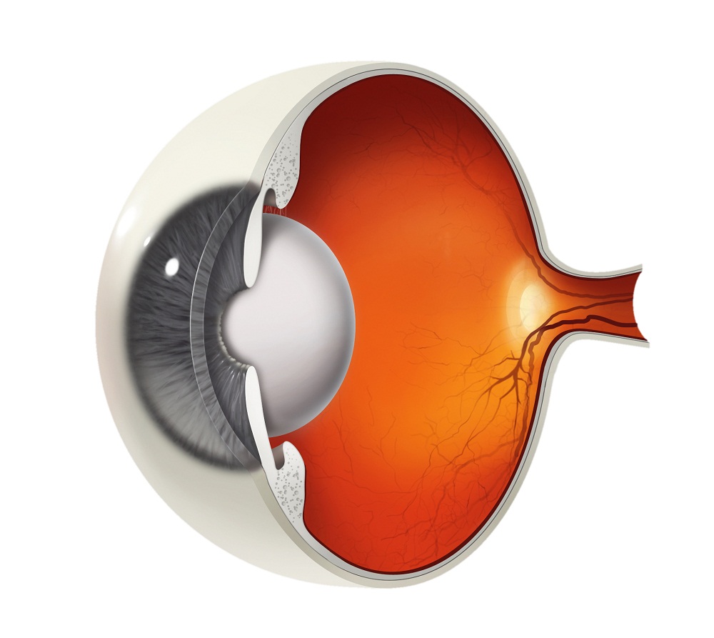

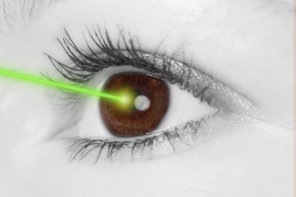

.jpg)
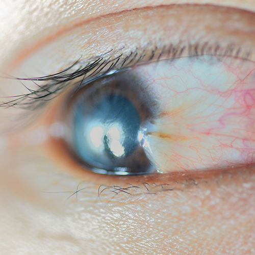
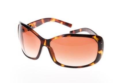


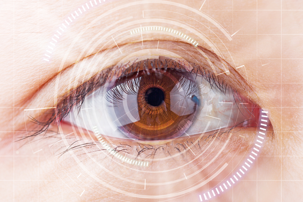
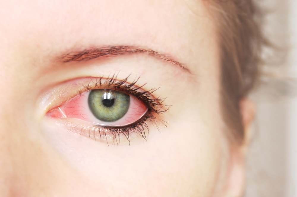
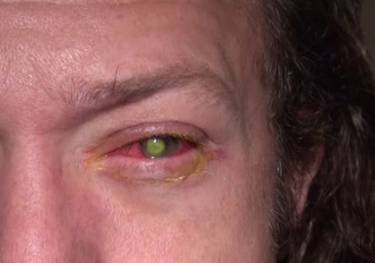

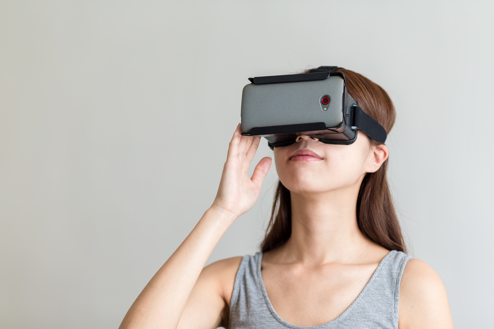
.jpg)

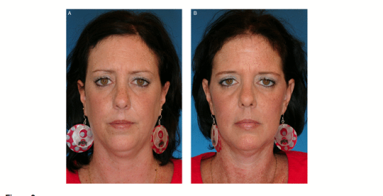
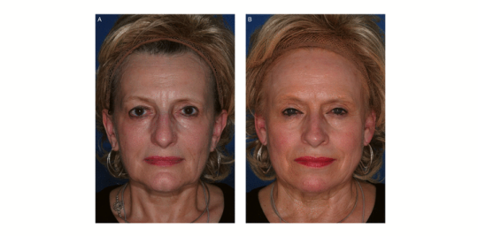
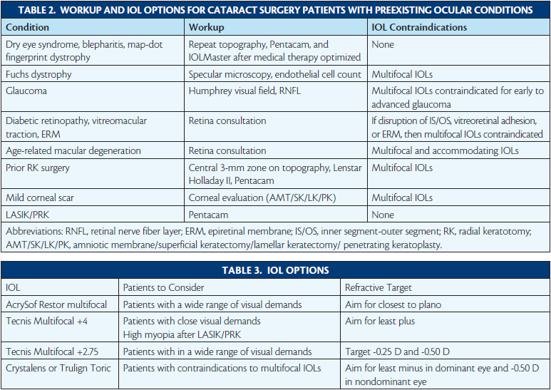


-page-001.jpg)
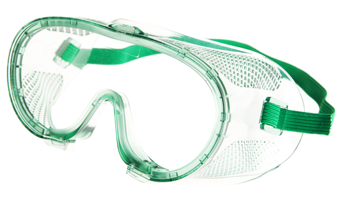
.jpg)
.jpg)


